baby chest x ray radiation
In general terms the risk to the unborn foetus from ionising radiation used for medical diagnosis X-rays CT nuclear medicine and angiography for example is dependent on. Long-term problems are very small.

Radiation From X Rays And Living In Denver They Re Not The Same
However radiation therapy has been shown to decrease milk production in some women and.

. 1317 years average 15 years. The most important downstream consequence specifically of X-rays for bronchiolitis is the inappropriate use of antibiotics Burstein says. These are plastic clips used to clamp the.
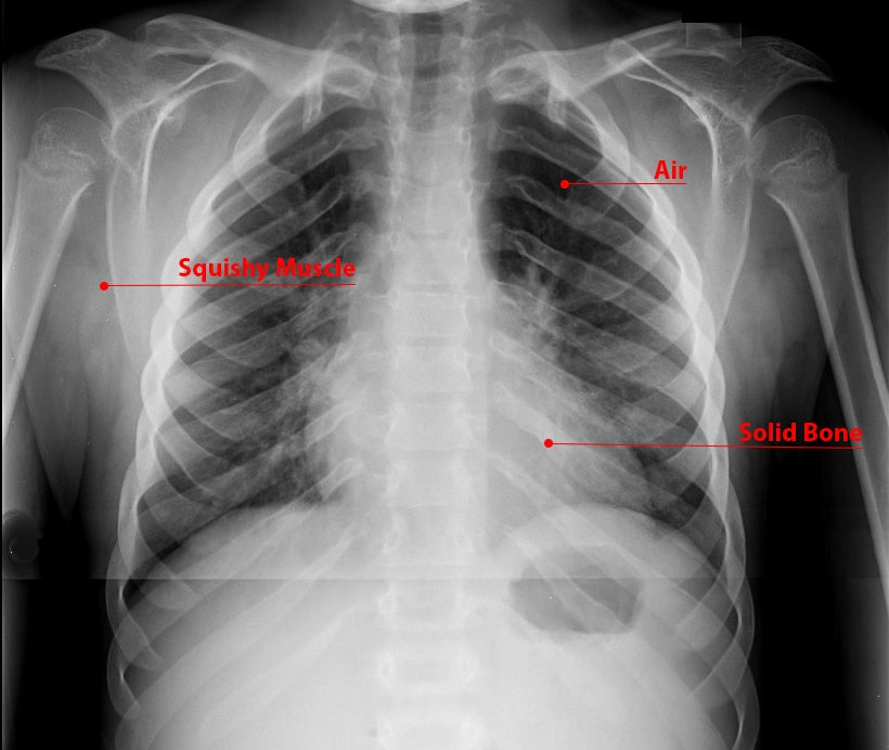
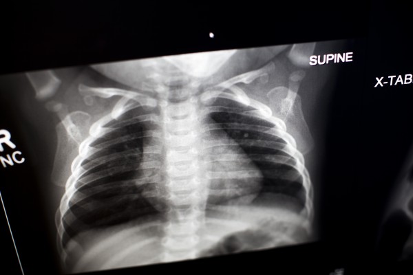
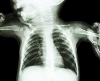
In a chest X-ray an X-ray machine sends a beam of radiation through the chest and an image is recorded on special film or a computer. An X-ray is a picture which is taken using a form of radiation that is able to pass through the body to create a digital X-ray image. This image includes organs and structures such as the heart lungs large blood vessels diaphragm part of the airway the upper spine ribs collarbone and breastbone.
The exception is abdominal X-rays which expose your belly and your baby to the direct X-ray beam. About one in. The part of the mothers body exposed to the radiation.
When the chest radiograph also includes the abdomen look out for the umbilical clip. Simple X-ray radiographs give very little radiation exposure. It is not putting your child at any risk.
An x-ray exam is a noninvasive medical test that helps doctors diagnose and treat medical conditions. One single chest x-ray is not concerning at all--even to newborns. Radiation exposure from X-rays does not pose any short-term problems.
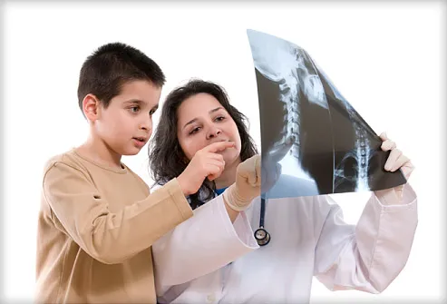
Risks depend on the amount of radiation to which the baby was exposed and the amount of time that it was exposed. When the X-rays pass through the body they create an image like a shadow. The X-rays are emitted from a special device with the child standing between it and a camera.
Depending on the patients age the difficulty of the examination will vary often requiring a specialist trained radiographer familiar with a variety of distraction and immobilization techniques. Whats a Chest X-Ray. Infants will need to be held.
Chest X-Ray CXR This test is performed to obtain an image of the heart lungs and ribcage. The most sensitive time period for central nervous system teratogenesis is between. Usually the X-ray technician will take pictures of the.
Fetal doses resulting from radiological examination of the mothers skull head neck chest and extremities are extremely low 001 rad because of the relatively low maternal radiation dose beam direction and distance between the primary field and the fetus. Chest PAAP erect 180 cm. 851a a Use automatic exposure control 500 speed for chestabdomen else 400 speed at specified kVp when practical.
The Alliance for Radiation Safety in Pediatric Imaging reminds parents and pediatricians to follow these guidelines. The child keeps as still as possible for a few seconds whilst the radiographer takes the picture. The FDA recommends that medical x-ray imaging exams which include computed tomography CT fluoroscopy and conventional X-rays use the.
1 A recent study claiming an association between dental radiography in pregnancy and low birth weight. Most X-ray exams including those of the legs head teeth or chest wont expose your reproductive organs to the direct X-ray beam and a lead apron can be worn to provide protection from radiation scatter. An x-ray exam may be performed on newborns infants and older children.
It is very rare for a single diagnostic x-ray to exceed even 5 rad. 33 rows For example the amount of exposure to the fetus from a two-view chest x-ray of the mother is only 000007 rad. Different parts of the body contain different tissues which vary in how much X-ray radiation they absorb depending on how dense they are.
37 years average 5 years. Diagnostic x-rays and other medical radiation procedures of the abdominal area also deserve extra attention during pregnancy. X-ray exams use a small dose of ionizing radiation to produce pictures of the inside of the body.
X-rays are the oldest and most frequently used form of medical imaging. This brochure is to help you understand the issues concerning x. Radiation exposure from X-rays may slightly raise the risk of later cancer especially in children who have had many tests with high radiation exposure.
Chest AP erect in chair 180 cm. 812 years average 10 years. The procedure is entirely painless.
First of all have a look to see if the neonate is premature or not - signs of prematurity being reduction in subcutaneous fat and the lack of humeral head ossification the latter occurs around term. It would take more than 50 chest x-rays to reach 5 rad. For example if the radiation dose to the unborn baby was roughly equivalent to 500 chest x-rays at one time the increase in lifetime cancer risk would be less than 2 above the normal lifetime cancer risk of 40 to 50.
According to the American College of Radiology no single diagnostic x-ray has a radiation dose significant enough to cause adverse effects in a developing embryo or fetus. If the mother is undergoing low dose radiation therapy on a localized area according to the British Journal of Radiology the mother can still choose to breastfeed if both the mother and babys doctor feel it is safe and no potential risk is posed to the infants over all health. You would have to x-ray your arm or leg more than 5000 times in order to reach 5 rad of exposure to your unborn baby.
The average child now gets seven scans that rely on radiation before age 18 one recent study shows. The age groups were based on exposures suitable for tissue thickness in the direction of the X-ray beam of a patient of averagestandard size in that age group for each projection. Radiation exposure from X-rays does not pose any short-term problems.
Long-term problems are very small. X-rays are often used to diagnose bone fractures and dislocated joints. Most of those tests are X-rays which use relatively low levels of radiation.
The chest radiograph is one of the most commonly requested radiographic examinations in the assessment of the pediatric patient. 901a a Use automatic exposure control 500 speed for chestabdomen else 400 speed at specified kVp when practical. Radiation exposure from X-rays may slightly raise the risk of later cancer especially in children who have had many tests with high radiation exposure.
Theres far less radiation exposure with an X-ray. Some common diagnostic procedures include dental chest CT scan headchest and abdominal view.

X Rays And Unshielded Infants Raise Alarms The New York Times

X Rays And Unshielded Infants Raise Alarms The New York Times

X Rays And Unshielded Infants Raise Alarms The New York Times

Estimation Of Radiation Dose During Diagnostic X Ray Examinations Of Newborn Babies And 1 Year Old Infants Semantic Scholar

Pem Pearls Chest Radiographs For Shortness Of Breath
What Is An X Ray For Kids Radiology And Medical Imaging

Neonatal Chest Radiography Influence Of Standard Clinical Protocols And Radiographic Equipment On Pathology Visibility And Radiation Dose Using A Neonatal Chest Phantom Radiography
X Ray For Kids Children S Health Orange County

Children S National Nicu Reduces Chest X Rays Unintended Extubations Innovation District Children S National

Chest X Rays For Children Doctors Are Unnecessarily Exposing Kids To Radiation With No Clinical Benefit

Radiation Doses In Neonatal X Ray Examinations Download Table
Lifetime Cancer Risks From X Rays For Children Relatively Low

Is A Single Chest X Ray Too Much Radiation For A 3 Year Old Healthychildren Org

Indications For Chest X Rays In Children And How To Obtain And Interpret Them

Ai Tool Uses Chest X Ray To Differentiate Worst Cases Of Covid 19 Imaging Technology News
Children S Radiation Exposure From X Rays And Ct Scans

Pediatric Chest Supine View Radiology Reference Article Radiopaedia Org

Pdf Radiation Protection In Pediatric Radiography Introducing Some Immobilization And Protection Equipment Semantic Scholar
